
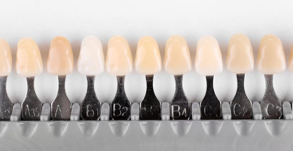

Digital shade matching replaces the subjective eyeball-and-tab method of choosing tooth colour with objective, reproducible data — from a spectrophotometer, an intraoral scanner, or calibrated cross-polarised photography — that can be sent straight to the lab. It matters because shade is one of the most common reasons an otherwise excellent anterior restoration comes back for a remake, and the fix is rarely at the bench: it is in how the colour was captured and communicated in the first place. This guide covers why visual shade-taking is unreliable, which digital methods work, and how to send colour the lab can actually reproduce.
Why visual shade-taking fails so often
Choosing a shade by holding a tab against a tooth is fast, but it is subject to everything the human visual system does badly: variable ambient lighting, metamerism, colour fatigue, dehydration of the tooth during the appointment, and simple inter-observer disagreement. Two clinicians — or the same clinician on two days — will often pick different tabs for the same tooth. The evidence bears this out: instrumental methods produce fewer colour errors and less incorrect shade matching than conventional shade tabs.¹² The problem is not the technician’s skill; it is the quality of the colour information they are given to work from.
The digital methods, and how they compare
Three tools dominate, and they are not equal:
- Spectrophotometer (e.g. intraoral units) — the most accurate and repeatable. Studies report a marked increase in accuracy and a more objective match in the large majority of cases compared with the human eye, and it consistently ranks highest for intra-instrument reliability.¹⁴
- Cross-polarised photography — a polarising filter cuts surface reflection so the true body colour and characterisation show. It is highly reproducible (repeated measurements differing by well under half a ΔE unit) and, with a grey reference card for calibration, reliable regardless of surgery lighting — and it gives the technician a map, not just a tab number.³⁴
- Intraoral scanner shade — convenient because it comes with the scan you are already taking, though it generally ranks a step below the spectrophotometer and cross-polarised photography for reliability.⁴
The practical hierarchy: spectrophotometer for the objective number, cross-polarised photography for the map and characterisation, scanner shade as a useful adjunct that arrives free with the digital impression.
How to send shade the lab can reproduce
Accurate capture is only half the job; communication is the other half. For an anterior aesthetic case, the lab benefits most from:
- An objective value — the spectrophotometer reading or scanner shade as a starting point.
- Cross-polarised photographs with a shade tab held in the same plane as the tooth and a grey card in frame for calibration.
- A shade map noting incisal translucency, cervical chroma, cracks, white spots and other characterisation — the detail a single tab can never carry.
- Photos taken before the tooth dehydrates, ideally early in the appointment.
Sent together from a clean digital impression, that package lets the lab match not just the base shade but the character that makes an anterior restoration disappear — the same attention to aesthetic detail behind cosmetic and anterior work.
Digital and visual together
Digital does not abolish clinical judgement. The most reliable results come from combining instrumental data with visual confirmation — the spectrophotometer or photograph gives an objective anchor, and the clinician’s eye validates it in context.² Treat the digital reading as the ground truth to communicate, and the visual check as the sanity test, rather than choosing one over the other.
How Zenith uses your shade data
Digital shade matching is part of Zenith’s workflow. Because the founder is a practising dentist, we know an aesthetic case is won or lost on the colour information we receive — so we work from your spectrophotometer values, cross-polarised photographs and shade maps to build the base shade and characterisation into the design, and confirm it against your scan. Across our restorative work we hold a 2% remake rate against an 8–12% industry average, and shade-related remakes are exactly the kind that good colour communication prevents. You can see how this feeds our crown, bridge and aesthetic restorations across the full range of services, and we accept scans from 3Shape, iTero, Medit, DS Core, Carestream and Smilecloud.
For an anterior or shade-critical case, get in touch and send us your shade records and we’ll match it with you.
FAQs
Frequently asked questions
Everything you need to know about working with Zenith Labs.
What is digital shade matching?
It is selecting tooth colour with instruments rather than the eye alone — using a spectrophotometer, intraoral scanner shade, or calibrated cross-polarised photography — to produce objective, reproducible colour data that can be communicated accurately to the lab.
How do I submit my first case?
Generally, yes. Instrumental methods produce fewer colour errors than conventional shade tabs, and spectrophotometers in particular show a marked accuracy improvement and a more objective match than the human eye in most cases.¹² Visual confirmation still adds value as a final check.
Spectrophotometer, photos or scanner — which is best?
For reliability, the spectrophotometer ranks highest, followed by cross-polarised photography, then intraoral scanner shade.⁴ In practice, use the spectrophotometer or scanner for an objective value and cross-polarised photographs for the map and characterisation.
How do I send shade to the lab?
Send an objective value (spectrophotometer or scanner shade), cross-polarised photographs with a shade tab in plane and a grey card for calibration, and a shade map noting translucency, chroma and characterisation — captured before the tooth dehydrates.³⁴
Do I still need to take a shade if I’m scanning?
Scanner shade is a useful adjunct, but for a shade-critical anterior case it is best supplemented with cross-polarised photography and, where available, a spectrophotometer reading, plus a characterisation map — the scan alone rarely captures the full aesthetic detail.⁴
Sources
- Wiley, Journal of Esthetic and Restorative Dentistry (Czigola et al., 2021) — “Comparing the effectiveness of shade measurement by intraoral scanner, digital spectrophotometer, and visual shade assessment” — https://onlinelibrary.wiley.com/doi/10.1111/jerd.12810
- PMC — “Novel Trends in Dental Color Match Using Different Shade Selection Methods: A Systematic Review and Meta-Analysis” — https://www.ncbi.nlm.nih.gov/pmc/articles/PMC8778907/
- PubMed — “The accuracy of dental shade matching using cross-polarization photography” — https://pubmed.ncbi.nlm.nih.gov/31840142/
- Wiley, Journal of Esthetic and Restorative Dentistry — “Reliability of Dental Shade Selection Methods: Agreement Among Spectrophotometer, Intraoral Scanner, and Cross-Polarization Photography” — https://onlinelibrary.wiley.com/doi/10.1111/jerd.13458